Detailed Human Tooth Cross-Section for Dental Education
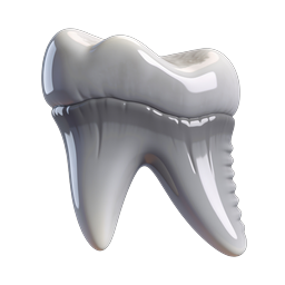
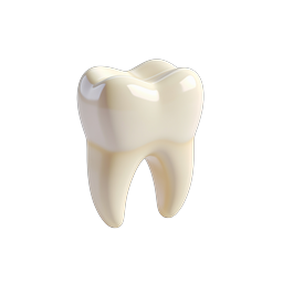
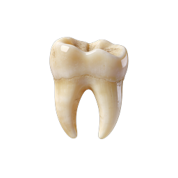
This detailed illustration represents a cross-section of a human tooth, showcasing its internal anatomical structures. The image displays the enamel (white outer layer), dentin (yellow layer beneath the enamel), pulp chamber (red innermost part containing nerves and blood vessels), and surrounding gum and bone tissues. Easily distinguishable layers highlight the complexity and essential functions of each part: enamel for protection, dentin for support, and pulp for sensory response and nourishment. This cross-sectional view is commonly used in dental education, research, and patient awareness materials to explain oral health, dental treatments such as root canal therapy, and common oral diseases like cavities and pulpitis. The use of realistic textures and accurate coloring enhances its value for dental professionals, students, and clinics aiming to illustrate the internal anatomy of teeth and the significance of maintaining oral hygiene.
- Format
- PNG · transparent
- Dimensions
- 256 × 256 px
- Use for
Image Generator Prompt
Free account required · 256 px free · Credit required · License