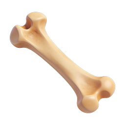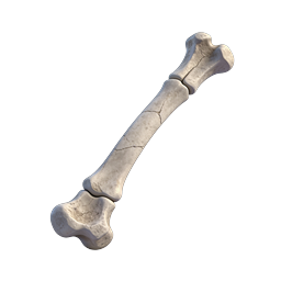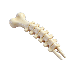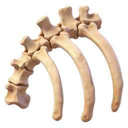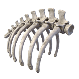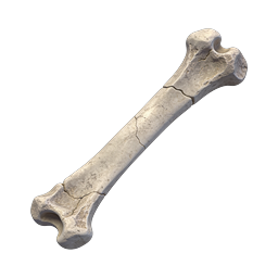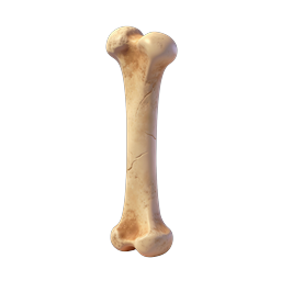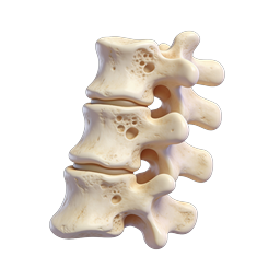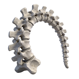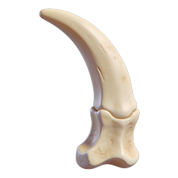
3D Knee Joint Anatomy Icon - Realistic Medical Illustration
This detailed 3D rendered icon depicts a human knee joint anatomy from a lateral side view, showcasing the intricate skeletal structure of the lower limb. The illustration features the distal femur (thigh bone) curving elegantly downward to articulate with the proximal tibia (shin bone), with visible articular cartilage lining the joint surfaces. The patella (kneecap) region is subtly indicated at the front of the joint, while the intercondylar notch and tibial plateau are clearly defined. The realistic bone texture includes subtle porous details, natural color variations in cream and beige tones, and smooth surface contours that highlight the anatomical accuracy. This icon is ideal for orthopedic websites, medical education platforms, physiotherapy apps, healthcare presentations, anatomy textbooks, surgical training materials, joint replacement informational content, sports medicine resources, and any digital application related to musculoskeletal health, bone biology, or medical visualization requiring precise anatomical reference imagery.
- Format
- PNG · transparent
- Dimensions
- 256 × 256 px
- Use for
Image Generator Prompt
Free account required · 256 px free · Credit required · License