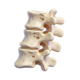
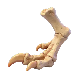
3D Foot Bone Anatomy Model - Skeletal Structure Icon
Detailed 3D rendered illustration of human foot skeletal anatomy, showcasing the intricate bone structure including the calcaneus (heel bone), talus, navicular, cuboid, cuneiforms, metatarsals, and phalanges. The realistic bone model features a natural cream and beige color palette with subtle textural details representing bone surface irregularities. Ideal for medical education platforms, orthopedic resources, podiatry clinics, anatomy textbooks, and healthcare presentations. This anatomical asset serves podiatrists, orthopedic surgeons, medical students, physical therapists, and fitness professionals teaching foot biomechanics. Perfect for use in medical apps, patient education materials, sports medicine content, dance and athletic training resources, and academic publications discussing foot pathology, fractures, arthritis, or surgical procedures.
- Format
- PNG · transparent
- Dimensions
- 256 × 256 px
- Use for
Image Generator Prompt
Free account required · 256 px free · Credit required · License